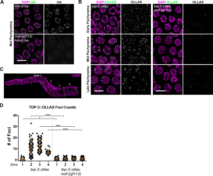
Fig 4. Localization of HIM-6 and TOP-3 in the rmif-2 mutant.
(A) Representative him-6::ha and rmif-2(jf113) him-6::ha nuclei in mid pachynema stained with DAPI (in magenta) and HA (in green). HIM-6 localizes to bright foci throughout pachynema in him-6::ha. In the rmif-2 mutant, HIM-6 is detected in small, faint foci throughout pachynema. Scale bar: 5 μm. (B) Representative images of nuclei throughout pachynema stained with DAPI (in magenta) and OLLAS (in green). TOP-3::OLLAS localizes to distinct foci throughout early, mid, and late pachynema. In the rmif-2 mutant, TOP-3 fails to localize properly, and only a few cytoplasmic and nuclear foci can be observed. Scale bars: 5 μm. (C) For the quantification of TOP-3::OLLAS foci three gonads per genotype were each divided into four equal zones from the transition zone (beginning of meiosis) until late pachynema. (D) Quantification of TOP-3 foci in top-3::ollas and top-3::ollas; rmif-2 backgrounds, throughout the C. elegans gonad. The mean number of TOP-3 foci in each zone was WT: zone 1: 0.4 (±0.8 SD), n = 97 nuclei; zone 2: 11.5 (±6 SD), n = 89; zone 3: 13.1 (±5 SD), n = 79; and zone 4: 6.8 (±1.6 SD), n = 57; rmif-2: zone 1: 0.6 (±0.9 SD), n = 103 nuclei; zone 2: 0.9 (±1.0 SD), n = 109; zone 3: 1.0 (±1.0 SD), n = 94; zone 4: 0.7 (±0.98 SD), n = 57.

