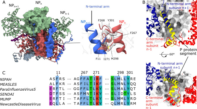
Fig 2. Protomer-protomer interactions within the NiV nucleocapsid assembly.
(A) Three adjacent protomers, where the two outside protomers are presented as ribbons and the central protomer is shown in surface representation, with the NT-arm in blue, N-terminal Ncore in green, C-terminal Ncore in coral and CT-arm in grey, as in Fig 1B. A magnified view of the molecular interaction of the NT-arm and the C-terminal Ncore domain is shown on the right with interacting residues (sticks) displayed within the CryoEM map. (B) The P protein segment from the structure of the complex with the monomeric NiV N form (yellow ribbon, pdb:4co6)[15] superimposed onto the central subunit in (A), shown in white surface representation. The two adjacent protomers are presented as dark-blue (n+1) and red (n-1) ribbons. (C) Alignment of interacting residue segments, shown in the magnified view in (A), for N proteins from several Paramyxoviruses, with conserved residues highlighted using the ClustalX colour scheme.

