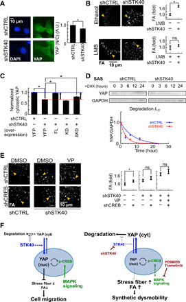
Fig. 5. STK40 and MAPK signaling regulated YAP-mediated FA remodeling in distinct mechanisms.

(A) Representative images and quantification of subcellular YAP distribution. Notice the decreased N/C ratio of YAP by shSTK40. A.U., arbitrary units. (B) Representative images and quantification of FA in SAS cells without or with leptomycin B (LMB) treated with shCTRL or shSTK40. Note the vanishment of STK40 effect under leptomycin B. (C) SAS cells received STK40 knockdown (shSTK40) followed by rescue experiments using STK40 of FL, KD, and ΔKD constructs. Their cytosolic YAP levels were measured using immunofluorescence. Notice that FL and ΔKD rescued YAP levels that were reduced by shSTK40. shCTRL, control shRNA; YFP, control vector. (D) Top: SAS cells with STK40 knockdown (shSTK40) were treated with cycloheximide. Their YAP protein was collected at different time points after cycloheximide treatment and measured using Western blots. Bottom: Quantification of the above results. Note that shSTK40 facilitated YAP degradation. (E) Representative images and quantification of FA in SAS cells with shSTK40 and treated with VP and/or shCREB. Note that shSTK40 and VP abolished effects of shCREB. Error bars denote means ± SEM. *P < 0.05. (F) Working models depict how STK40 and MAPK collaboratively regulate YAP activities (left) and how STK40 plus MAPK suppression causes synthetic dysmobility (right).
