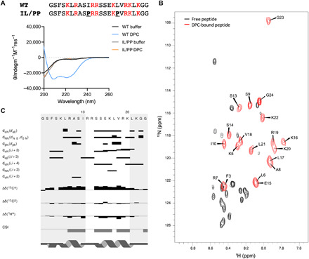
Fig. 2. The AH peptide folds into a helix in the presence of model membranes.

(A) Peptide sequences and their CD spectra in the absence or presence of 0.35% DPC. (B) Overlaid 15N HSQCs of the unbound (black) and DPC-bound (red with assignments) peptides. ppm, parts per million. (C) NOE/chemical shift index (CSI) plot of the DPC-bound peptide sequence.
