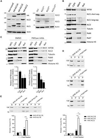
Fig. 5. Membrane association of kinesin-1 is mediated by KLCCTD and is phosphoinositide sensitive.

(A) KLC1/2 expression in HeLa cells transfected with HA-KLC1A/1E/2 or nontransfected cells (left) or after siRNA knockdown of KLC1/2 (right) was analyzed by Western blot. (B) Fractionation and Western blot analysis of endogenous KIF5B and KLC1/2. (C) Fractionation analysis of cells treated with dimethyl sulfoxide (DMSO) or PIKfyve inhibitor YM201636 (10 μM, 4 hours). Means ± SEM of at least three experiments. *P < 0.05, **P < 0.01, and ***P < 0.001 compared to the sample treated with DMSO. (D) Cosedimentation assay and analysis by SDS-PAGE/Commassie staining using purified KHC-KLC1B/1E/2 and Folch fraction I LUV. (E) Cosedimentation assay and analysis by SDS-PAGE/Commassie staining using purified KHC-KLC1E or –I/P KLC1E and Folch fraction I LUV. Means ± SEM of at least three experiments. **P < 0.01 and ***P < 0.001 compared to the sample without liposomes or as indicated in the graphs.
