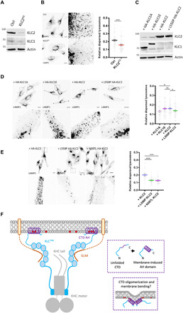
Fig. 6. Long KLCs are required for kinesin-1–mediated lysosome positioning.

(A) Western blot analysis of KLC2 expression in HeLa cells after siRNA knockdown of KLC2. (B) Lysosome distribution in control or KLC2 knockdown cells. Lysosome dispersion was determined by measuring perinuclear and whole-cell LAMP1 signal fluorescence. Means ± SEM of at least three experiments. ***P < 0.001. (C) KLC1/2 expression in KLC2 knockdown HeLa cells transfected with HA-KLC1A/1E/2/L550P 2 was analyzed by Western blot. (D) Lysosome distribution in KLC2 knockdown cells transfected with HA-KLC1A/1E/2/L550P 2. Means ± SEM with samples pooled from three independent experiments. *P < 0.05. (E) Lysosome distribution in KLC2 knockdown cells transfected with HA-KLC2 or N287L/L550P variants. Means ± SEM with samples pooled from three independent experiments. ***P < 0.001. (F) Model showing representation of kinesin-1 cargo membrane association by the KLCs. The membrane-induced helical folding and membrane remodeling capacity of the KLCCTD helix are also represented. ns, not significant.
