Figure 1.

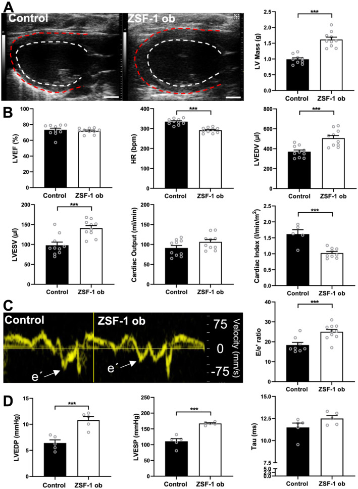
Echocardiographic and haemodynamic confirmation of HFpEF phenotype in ZSF‐1 obese rats. (A) Representative parasternal long‐axis B‐mode images of the left ventricle and quantification of left ventricular (LV) mass in grams (g). (B) Quantification of left ventricular ejection fraction (LVEF) in per cent (%), heart rate (HR) in beats per minute (b.p.m.), LV end‐diastolic and end‐systolic volumes in μL (LVEDV and LVESV), cardiac output (CO) in mL/min, and cardiac index (CI) in L/min/m2. (C) Representative tissue Doppler recordings from the septal mitral valve annulus depicted as velocity in mm/s and quantification of E/e′ ratio. (D) Haemodynamic data quantification of left ventricular end‐systolic/end‐diastolic pressures (LVESP and LVEDP) in mmHg as well as Tau in ms. n = 10 per group for echocardiography (except for CI: Control n = 5) and n = 5 per group for haemodynamics. Control, Wistar Kyoto rats; ZSF‐1 ob, ZSF‐1 obese. Scale bar = 3 mm; data are presented as mean ± standard error of the mean; statistics were performed by Mann–Whitney test or unpaired t‐test with *P < 0.05, **P < 0.01, ***P < 0.005.
