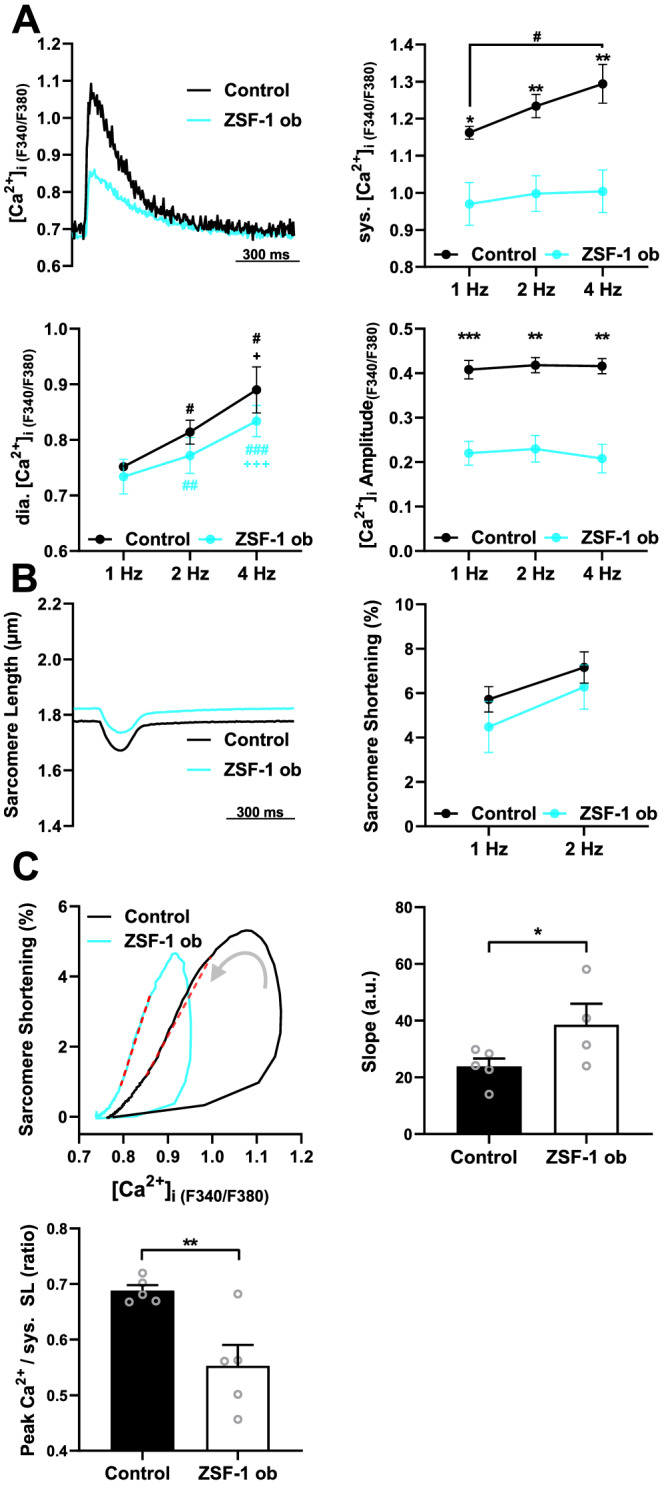
Figure 4.

Isolated right ventricular cardiomyocytes from HFpEF rats exhibit low intracellular [Ca2+] and preserved contractility. (A) Representative Ca2+ transients at 1 Hz (left, top), and mean values for systolic (sys.) and diastolic (dia.) [Ca2+], and Ca2+ amplitudes at 1, 2, and 4 Hz field stimulation. (B) Representative example of sarcomere shortening at 1 Hz (left) and mean sarcomere shortening amplitude (right) at 1 and 2 Hz. (C) Representative loops of sarcomere shortening as a function of [Ca2+]i in an RV cardiomyocyte from ZSF‐1 obese and Control rat (left). The linear fit between 20% and 80% relaxation is marked; mean values for the slope of the linear fit (right) and peak [Ca2+]i to syst. SL ratio (bottom); N = 4–5 animals per group (symbols) with an average of 16 cells per animal. Control, Wistar Kyoto rats; ZSF‐1 ob, ZSF‐1 obese. Error bars represent standard error of the mean; statistics were performed by two‐way analysis of variance with Fisher's least significant difference for (A) and (B) and unpaired t‐test for (C) with *P < 0.05, **P < 0.01, ***P < 0.005. # and + in (A) indicate differences compared with 1 and 2 Hz, respectively, within groups.
