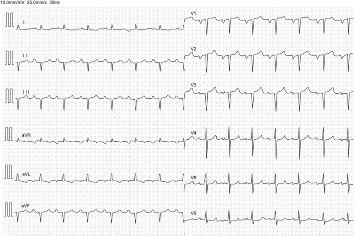
Figure 2.

Representative ECG image of a patient with ATTRwt‐CM. ECG showing QS pattern in the right pre‐cordial leads, left atrial loading, and cardiac conduction system disorder, including first‐degree atrioventricular block and left anterior fascicular block. Low voltage in limb leads on ECG may be less common at an early stage of ATTRwt‐CM. ATTRwt‐CM, wild‐type transthyretin amyloid cardiomyopathy; ECG, electrocardiogram.
