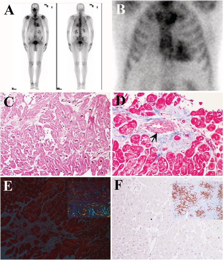
Figure 2.

Bone scintigraphy and LV Endomyocardial biopsy findings. (A, B) Whole body (A) and planar (B) images of the chest 90 minutes after 360 MBq of 99mTc diphosphonates i.v. administration, showing an evident accumulation of the radiopharmaceutical in the cardiac region (Perugini score 3). (C–F) LV endomyocardial biopsy revealing the presence of severely hypertrophied and disarrayed cardiomyocytes (C, haematoxylin and eosin, 200×), interrupted in short runs by interstitial and replacement fibrosis, with severe lumen narrowing of a small artery due to hypertrophy and hyperplasia of smooth muscle cells (D) (Masson thrichrome, 200×). LV endomyocardial biopsy did not show positivity either for apple‐green florescence at Congo‐Red staining (E, positive control in the square) or for TTR immunostaining (F, positive control in the square).
