. 2021 Jul 19;10:e66657. doi: 10.7554/eLife.66657

Figure 5. Three-dimensional structure of GloB.

(A) Overall fold, a-alpha helices colored in green and β-strands colored in purple. (B) Comparison of SaGloB (green) and human GloB (gray). (C) Docking of the substrate 1O (sticks) in the active site of GloB. Left, partial cartoon view; right, surface view. White represents hydrophilic residues, whereas red represents hydrophobic residues. Zn ions indicated as silver spheres; water indicated as blue sphere.

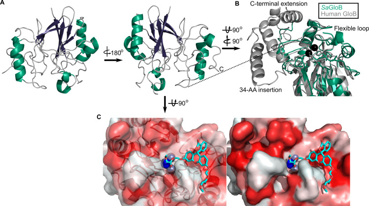
Figure 5—source data 1. Summary of crystallographic data collection and refinement statistics.

Figure 5.

Figure 5—figure supplement 1. Structural conservation of GloB.

Figure 5—figure supplement 1.

(A) Overall structural alignment of GloB (green) with S. enterica YcbI (PDB:2XF4), T. thermophilus TTHA1623 (PDB:2ZWR), and A. thaliana glyoxalase II (PDB:1XM8). Zinc coordinating residues are colored in green spheres. (B) Positioning of the zinc coordinating residues (green spheres).