. 2021 Jul 19;10:e68603. doi: 10.7554/eLife.68603

Figure 9. Pharmacological inhibition of ferroptotic stress blunts the accumulation of damage-associated PT cells and cell death.

(A) Schematic representation of experimental workflow. All mice (cKO and control littermates) were subjected to the same ischemic stress (ischemic time, 22 min, unilateral IRI) and tamoxifen treatment. The same volume of vehicle was administered to the control groups (control vehicle and cKO vehicle). Kidneys were harvested on day 21 post-IRI. (B) Liproxstatin-1 prevents renal atrophy. Relative size of post-IRI kidneys compared to contralateral kidneys (CLK) was quantified. Control, littermate control. N = 4–5. (C and D) Immunostaining for KIM1 and KRT8. IRI kidneys from cKO are shown. Quantification of immunostained area over the DAPI+ area is shown in (D). N = 4–5. (E and F) Immunostaining for SOX9 and VCAM1. Quantification of VCAM1+EMCNF4/80 area over the DAPI+ area is shown in (F). Arrowheads indicate damage-associated PT cells. (G) Real-time PCR analyses of indicated gene expression. Whole kidney lysates were used. N = 4–5. (H) and (I) TUNEL staining for evaluating cell death. Quantification of TUNEL-positive nuclei is shown in (I). N = 4–5. Red arrowheads indicate TUNEL+ tubular epithelial cells. Scale bars, 50 μm in (C) and (E); and 20 μm in (H). *p < 0.05; **p < 0.01; ***p < 0.001; ****p < 0.0001, unpaired t-test for (D), (F). and (I); One-way ANOVA with post hoc multiple comparisons test for (G). (J) Liproxstatin-1 improves renal repair after IRI.

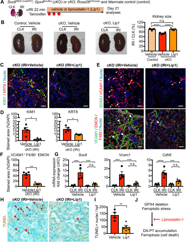
Figure 9.

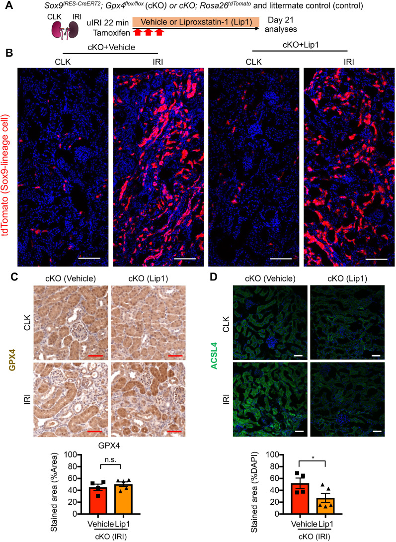
Figure 9—figure supplement 1. Liproxstatin-1 potently reduced ferroptotic stress in the absence of GPX4.

Figure 9—figure supplement 1.

(A) Experimental workflow for liproxstatin-1 (Lip-1) treatment to Gpx4 cKO mice. cKO mice and their littermate controls were subjected to the same ischemic stress (ischemic time, 22 min) and tamoxifen treatment. The same volume of vehicle was administered to cKO mice and littermate Cre-negative mice. Kidneys were harvested on day 21 post-IRI. (B) Distribution of tdTomato-expressing cells (Sox9-lineage cells) in contralateral (CLK) and IRI kidneys on day 21. Note that liproxstatin-1-treated kidneys and vehicle-treated kidneys show comparable targeting (n=three for vehicle, and n=two for liproxstatin-1). (C) Immunostaining for GPX4 and quantification. GPX4 immunostaining confirms the deletion of GPX4 in IRI-kidneys from cKO mice both vehicle and liproxstatin-1-treated group. N = 4–5. (D) Immunostaining for ACSL4 and quantification using kidneys from cKO mice (day 21 post-IRI). Liproxstatin-1 treatment reduced the expression of ACSL4 in IRI kidneys in the absence of GPX4. p<0.05, unpaired t-test. Scale bars, 100 μm in (B) and 50 μm in (C) and (D).
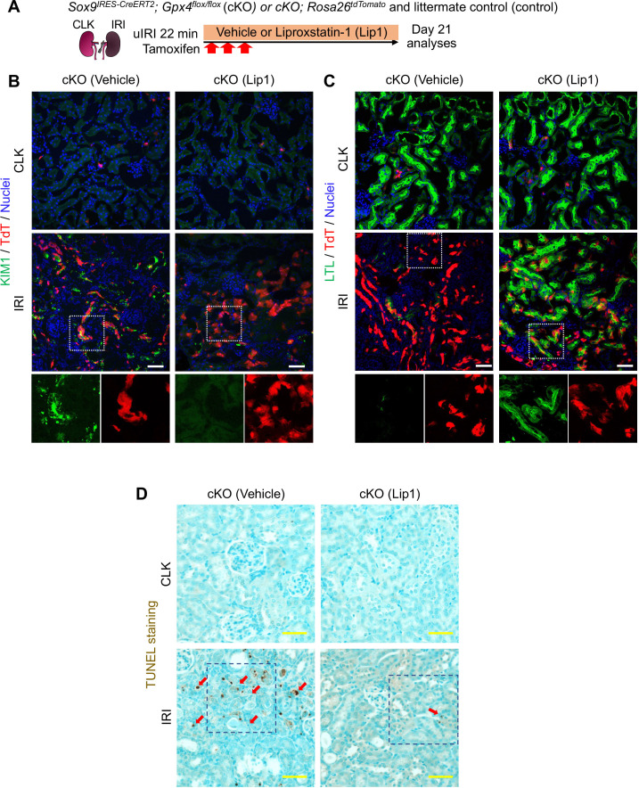
Figure 9—figure supplement 2. Liproxstatin-1 potently mitigated ferroptotic stress-induced pathologic changes in the absence of GPX4.

Figure 9—figure supplement 2.

(A) Experimental workflow for liproxstatin-1 (Lip-1) treatment to Gpx4 cKO mice. cKO mice and their littermate controls were subjected to the same ischemic stress (ischemic time, 22 min) and tamoxifen treatment. The same volume of vehicle was administered to cKO mice and littermate Cre-negative mice. Kidneys were harvested on day 21 post-IRI. IRI, ischemia-reperfusion-injured kidneys. CLK, contralateral kidneys. (B) and (C) Immunostaining for KIM1 and LTL in Sox9-lineage-tagged kidneys. Sox9-lineage cells express native tdTomato red fluorescence (TdT). Note that liproxstatin-1 treatment potently reduced the expression of KIM1 and restored the expression of LTL in tdTomato-positive cells in ischemia-reperfusion-injured cKO kidneys. Insets: individual fluorescence channels. Note that Inlets show the higher magnification and single-color images of dotted areas. (D) TUNEL staining for evaluating cell death. The dotted areas are shown in Figure 9H. See quantification for Figure 9I. N=4–5. Arrows, TUNEL+ nuclei. Scale bars, 50 μm in (B) – (D).