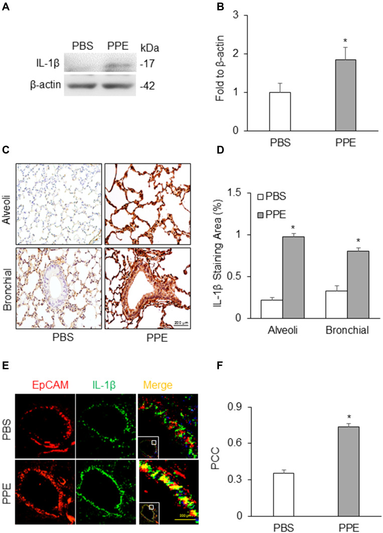
Figure 3.
NLRP3 inflammasome activation in the lung. (A) Semiquantitative immunoblots reacted with anti-IL-1β antibodies. β-actin was used as internal loading control. (B) Corresponding densitometric analyses of protein expression levels of cleaved IL-1β normalized by β-actin (n=3). (C) Representative micrographs depict IL-1β immunostaining in the alveolar and bronchial wall of lung. (D) Summarized bar graphs shows PPE significantly increased IL-1β immunostaining. (E) Representative micrographs depict colocalization of EpCAM and IL-1β in the bronchial epithelium in the lung. (F) Bar graph shows PCC of EpCAM and IL-1β significantly increased in the lung of mice receiving PPE instillation (n=5–7). *P<0.05 vs PBS treatment.

