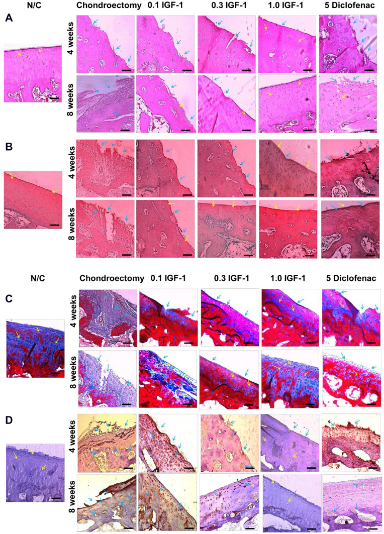
Figure 7.
Effects of IGF-1 on (A) hematoxylin-eosin staining (B) safranin O (C) masson’s trichrome staining for the existence of the cartilage during the OA in rabbit femoral condyle after treatment of at 4 and 8 weeks and (D) immunohistochemistry staining for the existence of MMP-1 in the cartilage at 4 and 8 weeks in vivo (× 200, scale bar = 200 μm). Yellow arrow indicated chondrocyte cell and blue arrow indicated erosion of condyle and positive MMP-1 (IHC).

