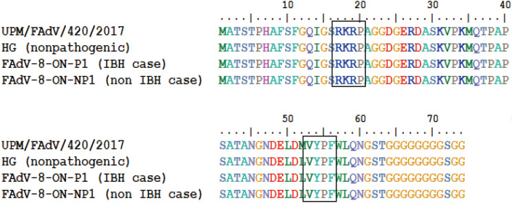
Fig. 3. The amino acid pairwise alignment of the fiber tail region as computed by ClustalW for comparison between studied isolate with reference isolate of non-pathogenic FAdV-8b and reference isolates from IBH and non-IBH cases [11]. The boxes indicate the presence of conserved sequence motifs found in the tail region of FAdV-8b.
FAdV, fowl adenovirus; IBH, inclusion body hepatitis.

