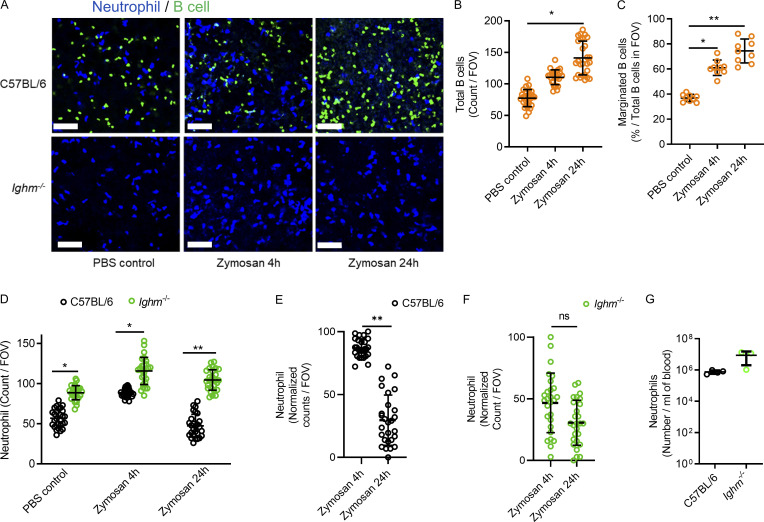
Figure 6.
Marginated B cells increase during direct zymosan-induced lung injury and restrain excessive neutrophil recruitment.(A) Lung intravital microscopy was performed on either C57BL/6 or Ighm−/− mice that were treated with PBS i.t. as a control or with 106 zymosan-coated beads i.t. Neutrophils and B cells were visualized using i.v. fluorescently conjugated anti-Ly6G mAb and fluorescently conjugated anti-CD19 mAb. Scale bar, 70 µm. (B and C) Total visualized B cells and marginated B cells were quantified in C57BL/6 mice, and no B cells were visualized in Ighm−/− mice. (D–F) Neutrophils were quantified and compared between mice that received PBS control or 4 h or 24 h of zymosan i.t. (G) Peripheral blood neutrophil counts were compared. For A–G, n = 3 independent experiments using nine mice in total. All individual imaging FOV replicates are shown for B–F. B and C were analyzed using one-way ANOVA and Tukey’s post hoc test. D–G were analyzed using Student’s t test based on n. Pooled data are presented as mean of n ± SD. *, P < 0.05; **, P < 0.01.

