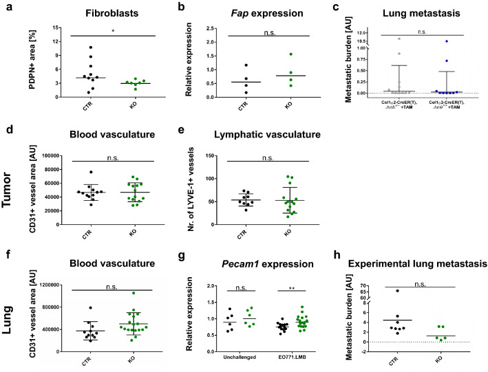
Fig. 3.
JUNB does not alter fibroblast density or tumor vasculature. a, b Quantification of fibroblast content in primary tumors by immunohistochemistry for Podoplanin (a; PDPN), n = 10 (CTR) and n = 7 (KO), Mann Whitney analysis: p = 0.0431, and gene expression analysis of fibroblast activating protein (B; FAP), n = 4 for both CTR and KO, Mann Whitney: p = 0.6857. c Distant lung metastasis in conditional fibroblast-specific mice as quantified via the presence of the mCherry reporter in tumor cells in whole genomic DNA, n = 11 (Col1α2-CreER(T), Junb+/+, + TAM) and n = 9 (Col1α2-CreER(T), Junb>/>, + TAM), data obtained from three independent injection rounds, Mann Whitney: p = 0.4119. d Tumor blood vasculature as assessed by CD31 immunofluorescence staining. Quantification was based on two whole tumor sections with 5 random fields each. Necrotic areas were avoided. Significance was assessed by unpaired t-test, p = 0.9521, n = 12 (CTR) and n = 14 (KO). e Number of lymphatic vessels in primary tumors, LYVE-1 + structures were manually counted on two whole tumor sections, n = 9 (CTR), n = 14 (KO). Unpaired t-test, p = 0.9449. f Blood vasculature in early metastatic lungs quantified by immunofluorescence staining for CD31, n = 11 (CTR), n = 18 (KO), unpaired t-test: p = 0.0895. g Gene expression analysis of Pecam1 in whole lungs of unchallenged and tumor-bearing mice, n = 6 (unchallenged CTR and KO), n = 14 (tumor-bearing CTR), n = 18 (tumor-bearing KO). Mann Whitney analysis of lungs from unchallenged (p = 0.3939) and tumor-bearing mice (p = 0.0079). h Experimental lung metastasis of EO771.LMB-mCherry cells as quantified by qPCR of the mCherry reporter on DNA level, n = 8 (CTR) and n = 5 (KO) from two independent injection rounds. Significance assessed by Mann Whitney test, p = 0.1709. *p < 0.05, **p < 0.01. Data are shown as means and SD in (d–f), in (a, b, g, h) the geometric mean and in (c) the geometric mean plus geometric SD are indicated. Data points represent individual mice

