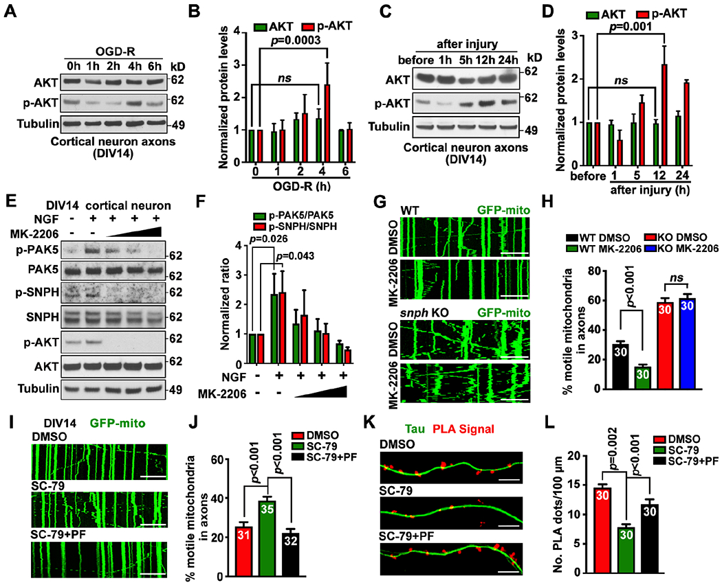
Figure 5. AKT signaling activates PAK5.

(A-D) Axonal AKT activation upon OGD-R (A, B) or axotomy (C, D) in microfluidic devices. Axoplasmic lysates were collected from axon chambers for immunoblotting. The intensity of AKT and p-AKT was quantified (n=3) and normalized to the 0-hour of OGD-R or before injury.
(E and F) AKT activates PAK5-SNPH signaling. Neurons (DIV14) were treated with NGF (40 ng/ml) and/or an increasing dose of AKT inhibitor MK-2206 (2, 4, or 6 μM) for 1 hour. Cell lysates (8 μg) were immunoblotted. The intensity ratios of p-PAK5/PAK5 and p-SNPH/SNPH were quantified (n=3) and normalized to the ratios in the untreated group.
(G and H) AKT-dependent remobilization of axonal mitochondria. WT or snph KO neurons were transfected with GFP-Mito at DIV10, followed by treatment at DIV14 with DMSO or AKT inhibitor MK-2206 (2 μM for 1 hour) before time-lapse imaging (90 frames with 5-sec intervals).
(I and J) Inhibiting PAK5 abolishes AKT-enhanced axonal mitochondrial transport. Neurons were transfected with GFP-Mito at DIV10, and treated with DMSO, AKT activator SC-79 (2 μg/ml), and/or PAK inhibitor PF-3758309 (PF, 1 μM) for 1 hour at DIV14, followed by time-lapse imaging (90 frames with 5-sec intervals).
(K and L) Activating AKT reduces in situ SNPH-MT association through PAK5. Neurons at DIV10 were treated with AKT activator SC-79 and/or PAK inhibitor PF-3758309 before PLA.
Data were quantified from n=30-40 neurons (H, J, L) per condition as indicated within bars from three experiments, expressed as mean±SEM, and analyzed by Student’s t test (H) or one-way ANONA with Dunnett’s multiple comparisons test (B, D, F, J, L). Scale bars, 10 μm.
