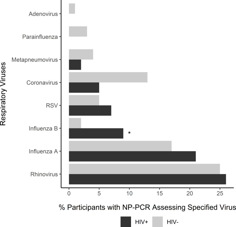
Figure 1.
Distribution of respiratory viruses isolated by RT-PCR from NP swabs among subjects with ILI, categorized according to HIV positivity. The bars represent the percentage of participants with a detected respiratory virus among those who had a documented NP PCR specimen result for any of the specified viruses. Multiple viruses may have been identified from a single sample. The number of sample results varied by virus (adenovirus, n=502; parainfluenza virus, n=469; metapneumovirus, n=469; RSV, n=469; coronavirus, n=425; influenza, n=503; and rhinovirus, n=489). *Statistically significant at a p value of <0.05, Fisher’s exact test. ILI, influenza-like illness; NP, nasopharyngeal; RSV, respiratory syncytial virus; RT-PCR, reverse transcription–PCR.

