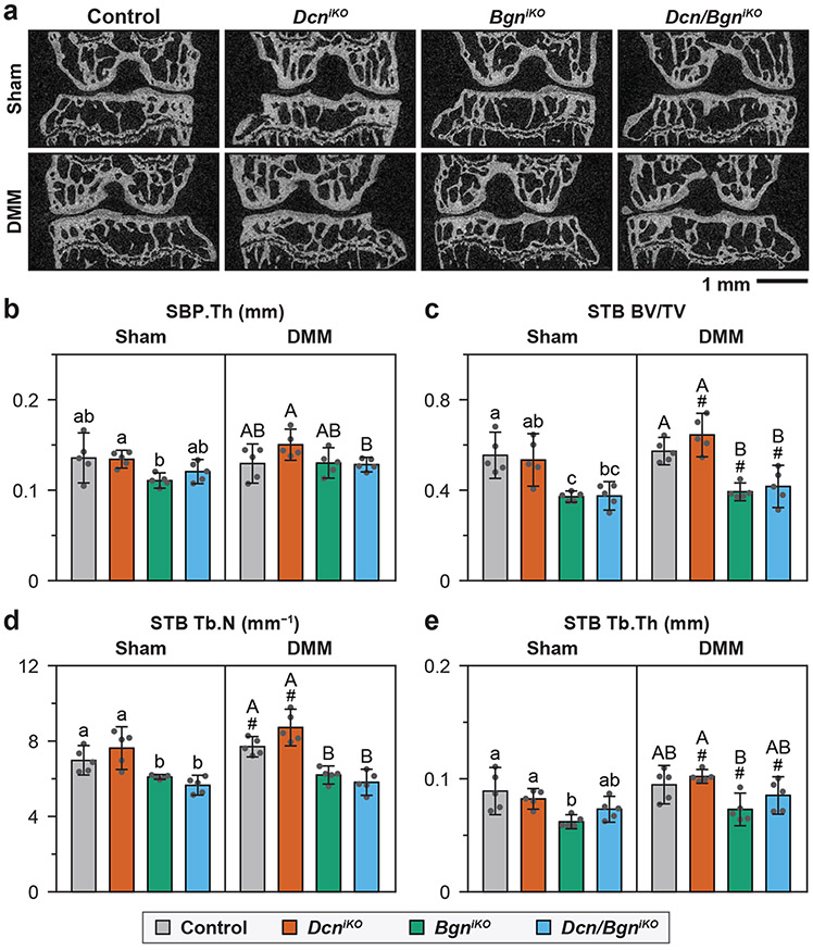
Figure 6.
a) Representative 2D μCT frontal plane images of the knee joint at 8 weeks after Sham and DMM surgeries (L: lateral, M: medial). b) Subchondral bone plate thickness (SBP.Th) and c-e) Subchondral trabecular bone structural parameters, including c) BV/TV: bone volume fraction, d) Tb.N: trabecular number, and e) Tb.Th: trabecular thickness, as measured from the μCT images of medial tibia. Panels b-e: mean ± 95% CI, n = 5, each data point represents the averaged value measured from one animal. Different letters indicate significant differences between genotypes for each surgery type, #: p < 0.05 between Sham and DMM surgeries for each genotype. Data for control and DcniKO mice are adapted from Ref. 15 with permission.

