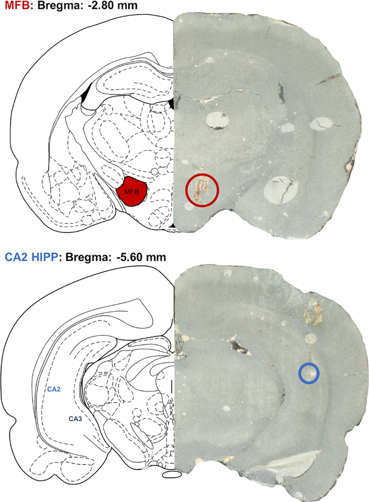
Fig. 3. Tissue analysis of FSCV electrode placement.

A graphic showing stimulating electrode placement in the MFB (top panel) and working electrode placement in the CA2 region of the hippocampus (bottom panel).

A graphic showing stimulating electrode placement in the MFB (top panel) and working electrode placement in the CA2 region of the hippocampus (bottom panel).