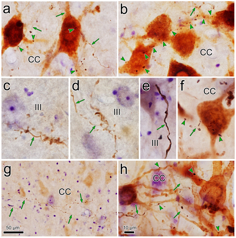
Figure 6.
Comparison of BDA labeled axonal arbors from different cases. a-b. Close associations (arrowheads) between BDA labeled axonal boutons and retrogradely labeled levator motoneurons in CC from a case with a pV injection that also involved the vestibular nuclei (Case 2). Most of the labeled axons (arrows) were quite fine in caliber. c-d. Examples of BDA labeled axons in III from the same case, showing grape-like organization of axon terminal arbors. e. An example of thick BDA labeled axon observed in III when the BDA spread into the vestibular nuclei (Case 5). f. Close associations between labeled boutons and labeled superior rectus motoneurons in a case where the pV injection spread into the vestibular nuclei (Case 3). g. Morphology of the fine BDA labeled axons in CC. (Case 2, but no reaction performed for WGA-HRP). h. Close associations between labeled boutons and labeled motoneurons in the case (Case 6) illustrated in figure 8. Scale in h=a-f.

