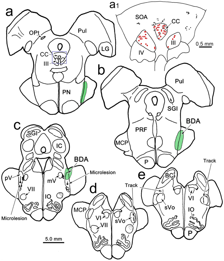
Figure 9.
Distribution of the peri-oculomotor terminal field following a physiologically localized (see Fig. 8) BDA injection (Case 6). Microlesions (c) and tracks (d-e) indicate areas in which neurons with receptive fields on the face were recorded in pV. This information was used to make a BDA injection in rostral pV (c), which spread further rostrally along the medial border of the middle cerebellar peduncle (MCP)(a-b). As shown in the higher magnification insert (a1), this injection produced terminal labeling (stipple) in SOA and CC. This animal also received a WGA-HRP injection in the levator and superior rectus muscles that retrogradely labeled motoneurons (dots) in III and CC.

