
Figure 4.
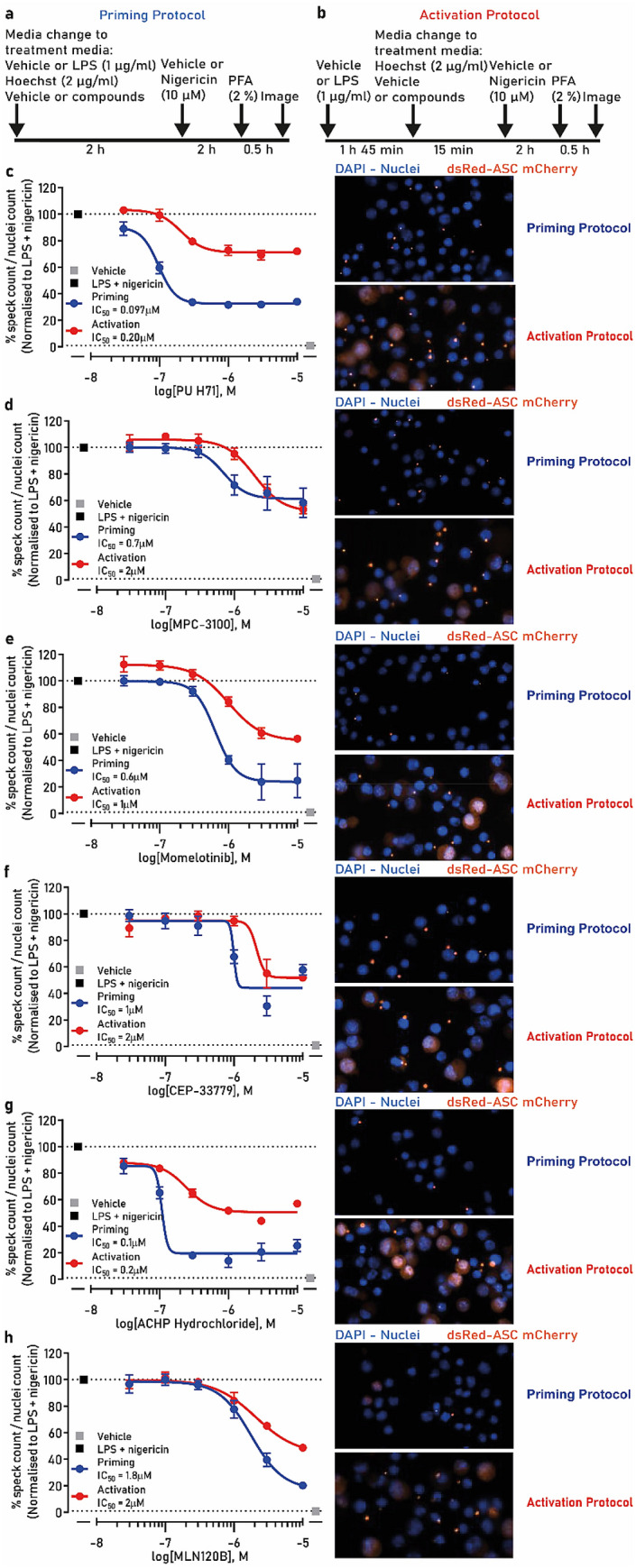
NLRP3 inflammasome inhibitor hits validation in the speck assay. (a) Schematic of the priming protocol. (b) Schematic of the activation protocol. ASC-mCherry iBMDM cells were treated with indicated concentrations of (c) PU H71, (d) MPC-3100, (e) momelotinib, (f) CEP-33779, (g) ACHP or (h) MLN120B with LPS (1 µg/ml) for 2 h followed by nigericin treatment (10 µM, 2 h) after which PFA was added (priming—blue trace) and specks counted. In a parallel experiment the compounds were added after LPS treatment just prior to nigericin (activation—red trace). The images are representative of the nuclei and speck formation for each compound at 10 µM in both priming and activation protocols. Data are presented as mean ± SEM, n = 3 independent experiments. Data in (c–h) was analyzed using GraphPad Prism version 7 software (https://www.graphpad.com/scientific-software/prism/).