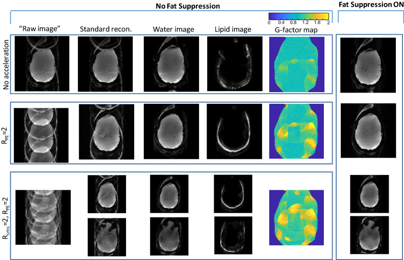
Figure 3.
Phantom scanning—combining in-plane acceleration, SMS and lipid-water separation. From top to bottom—Rtot = 2 no acceleration (Lipid/Water separation), Rtot = 4 (Lipid/Water, RPE = 2), Rtot = 8 (Lipid/Water, RPE = 2, Rsms = 2). From left to right—“raw image” (FFT applied directly to the acquired image), “standard recon.” (Siemens product reconstruction), images reconstructed with the extended formulation—separate water and lipid images, g-factor maps for the common slice, and the image acquired with fat suppression. The g-factor color-map range is 0–2.

