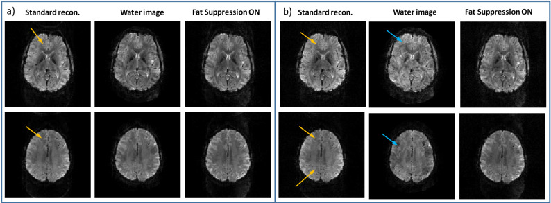
Figure 5.
Human imaging—combined in-plane acceleration, SMS and lipid-water reconstruction. Two representative slices are shown for (a) RPE = 2 and Rsms = 3 and (b) RPE = 2 and Rsms = 4. From left to right—standard recon. (Siemens product reconstruction), water image, and lipid image from scan without fat suppression, and the image acquired with fat suppression. Yellow arrows point to the artifact due to the lipid signal in the standard reconstruction. Blue arrows point to artifacts that occur due to high acceleration factor—Rtot.

