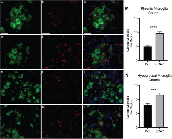
Fig. 4.
SCA7 mice exhibit increased amounts of activated microglia in the putative phrenic and hypoglossal nuclei. (A-L) Representative images of ChAT- (green), Iba1- (red) and DAPI- (blue) stained cervical spinal cord and medulla of 10-week-old wild-type (WT) and SCA7 mice. Putative phrenic motor nucleus of wild-type (A-C) and SCA7 (D-F) mice. Hypoglossal motor nucleus of wild-type (G-I) and SCA7 (J-L) mice. Quantification of Iba1+ cells revealed a significant increase in the number of microglia in putative phrenic (M) and hypoglossal (N) motor nuclei of SCA7 mice compared to wild-type mice. Data are mean±s.e.m. Statistical significance was determined using an unpaired two-tailed Student's t-test (***P<0.001, ****P<0.0001; phrenic, n=4 wild type, n=4 SCA7; hypoglossal, n=3 wild type, n=5 SCA7). Scale bars: 40 µm.

