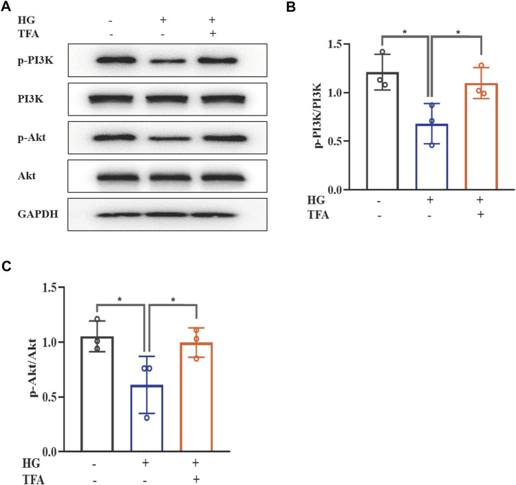
FIGURE 4.
TFA activated the PI3K/Akt signaling pathway in HG-afflicted MPC-5 cells. (A) WB analysis of p-PI3K, PI3K, p-Akt, and Akt, in cultured MPC-5 cells exposed to HG at 30 mM for 48 h with or without TFA (20 μg/ml) treatment for 24 h; (B) p-PI3K was quantified by densitometry; (C) p-Akt was quantified by densitometry. Data are expressed as mean ± S.D., (n = 3). *p < 0.05, **p < 0.01. Abbreviations: TFA, total flavones of Abelmoschus manihot; PI3K, phosphatidylinositol 3-kinases; Akt, protein kinase B; HG, high glucose; MPC-5, mouse podocyte cell-5; WB, western blotting; p-PI3K, phosphorylated-phosphatidylinositol 3-kinase; p-Akt, phosphorylated-protein kinase B.

