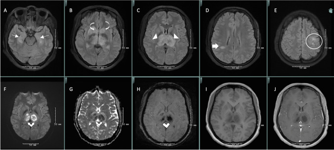
Fig. 1.
MRI brain was notable for T2-weighted-fluid-attenuated inversion recovery (T2-FLAIR) hyperintensities in bilateral thalami (white arrowheads) and caudate nuclei (curved white arrow) with hemorrhage (white double lines) and diffusion restriction (white double arrows), in addition to T2-FLAIR hyperintensities in bilateral hippocampi (thin white arrow), right parietal deep white matter (thick white arrow), and bilateral posterior frontal white matter (white circle), consistent with acute necrotizing encephalopathy due to SARS-CoV-2 (A–E axial T2-weighted-fluid-attenuated inversion recovery (FLAIR), F diffusion-weighted imaging, G apparent diffusion coefficient, H susceptibility-weighted imaging, I T1 pre-contrast, J T1 post-contrast)

