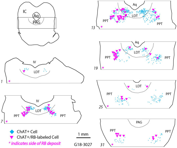
Figure 3.
Retrogradely labeled cholinergic cells (magenta triangles) were located in the pontomesencephalic nuclei ipsilateral and contralateral to an injection of red RetroBeads in the left SOC. Each symbol represents a single labeled cell. ChAT+ cells that did not contain RetroBeads are illustrated (cyan diamonds) to indicate the extent of the pedunculopontine and laterodorsal tegmental nuclei (PPT and LDT, respectively). Numbered sections are arranged from caudal to rostral and represent the dorsal tegmental region (indicated by the rectangle in the orientation section). The dashed line indicates the ventral border of the periaqueductal gray (PAG). The magenta asterisk at the bottom of each section outline indicates the side ipsilateral to the RB deposit in the SOC. Aq, cerebral aqueduct; IC, inferior colliculus, IV, fourth ventricle.

