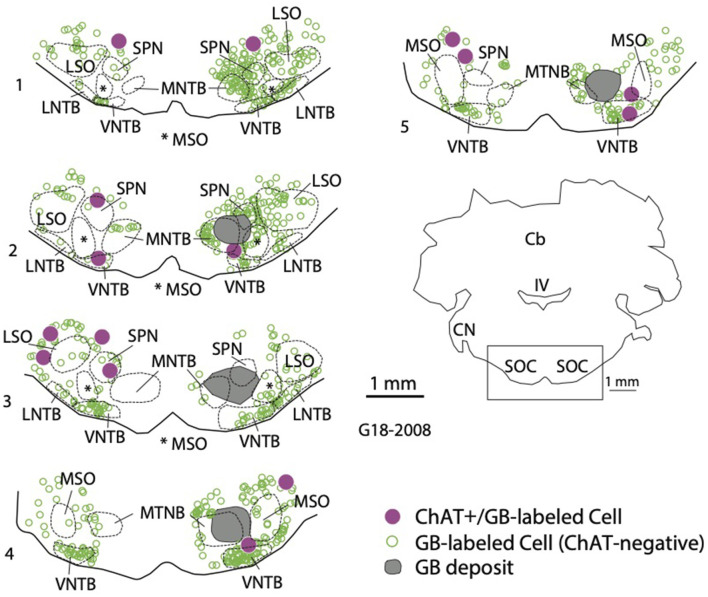
Figure 5.
Retrogradely labeled cholinergic cells were located in the SOC both ipsilateral and contralateral to a tracer deposit. The plot shows a deposit of green beads (GB, deposit shown in gray) in the right SOC. GB-labeled cells that were ChAT+ (magenta circles) were scattered among SOC nuclei on both sides. In addition, a large number of GB-labeled cells that were ChAT-negative were also labeled (open green circles). Numbered sections are arranged from caudal to rostral and represent the ventral portion of each section to show the SOC (indicated by the rectangle in the orientation section). IV, fourth ventricle; Cb, cerebellum; CN, cochlear nucleus; LNTB, lateral nucleus of the trapezoid body; LSO, lateral superior olivary nucleus; MNTB, medial nucleus of the trapezoid body; MSO, medial superior olivary nucleus; SOC, superior olivary complex; SPN, superior paraolivary nucleus; VNTB, ventral nucleus of the trapezoid body.

