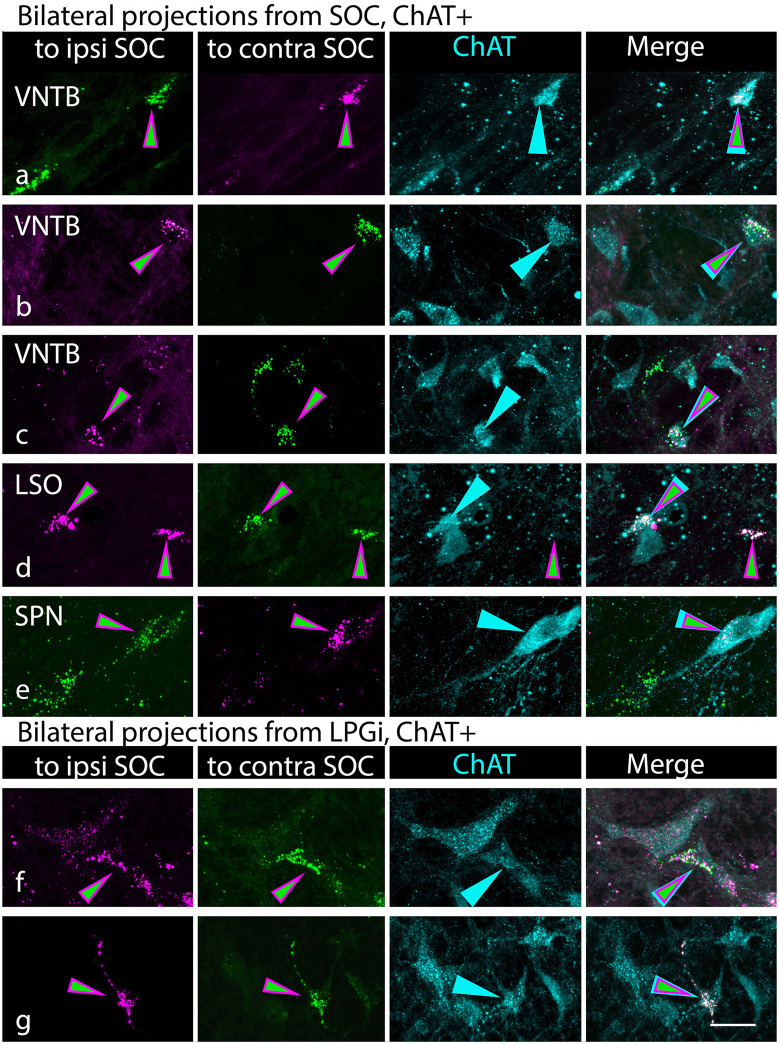
Figure 8.
Cholinergic cells in the superior olivary complex (SOC) and lateral paragigantocellular nucleus (LPGi) send branching axonal projections to innervate the SOC bilaterally. Each row of photographs shows a single field of view. The first two columns show the tracer label (RB in magenta; GB in green), with the first column showing the tracer injected into the ipsilateral SOC and the second column showing the tracer injected into the contralateral SOC (relative to the labeled cells). Magenta/green arrows identify cells that contain both retrograde tracers. Column 3 shows the ChAT staining, with cyan arrows pointing to the same cells as in the first two columns. Column 4 shows the merged image, highlighting the triple-labeled cells. (A–E) Triple-labeled cells were found in numerous SOC nuclei, indicated by the labels in column 1. VNTB, ventral nucleus of the trapezoid body; LSO, lateral superior olivary nucleus, SPN, superior paraolivary nucleus. ChAT-negative cells could also be labeled with both retrograde tracers (panel D, cell on the right). (F,G) Triple-labeled cells in the LPGi. Panels (A,B, and F) are from G18-2012 (medial SOC deposits); panels (C,D, and E) are from G18-3033 (lateral SOC deposits); panel (G) is from case G18-3030 (medial SOC deposits). Scale bar = 20 μm.

