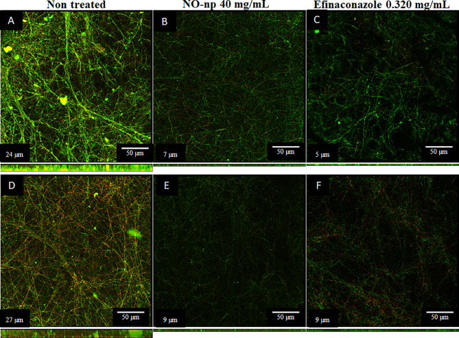
Figure 3.
Confocal microscopy images of mature T. rubrum biofilms formed on glass-bottom plates for 72 h at 37°C (A, D) and treated with NO-np (B, E) or EFCZ (C, F). Orthogonal images of mature T. rubrum biofilms showed metabolically active (red, FUN-1-stained) cells embedded in the polysaccharide extracellular material (green, ConA), while the yellow-brownish areas represent metabolically inactive or nonviable cells. Images were obtained after 72 h of exposure of the fungal cells to 40 mg/mL of NO-np or 0.32 mg/mL of EFCZ, and the images were compared with those of biofilms incubated in presence of RPMI. The pictures were taken at a magnification of ×63. Bars, 50 μm. The thickness of the fungal biofilms grown under these conditions was measured by z-stack reconstruction. The results are representative of those of two experiments.

