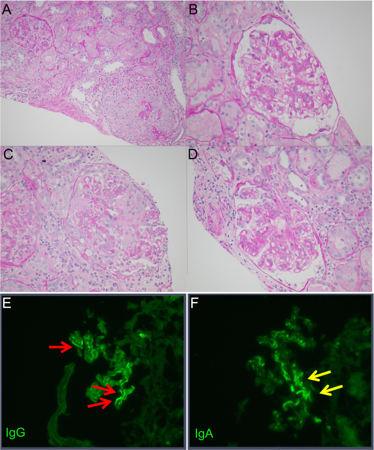
Figure 1.
Periodic acid-Schiff (PAS)-stained images of the kidney. (A) ×20 magnification of one glomerulus with fibrinoid necrosis, crescent and rupture of Bowman’s capsule and adjacent glomerulus with mildly hypercellular mesangium. (B) ×40 magnification of glomerulus with mild mesangial hypercellularity and mildly increased mesangial matrix. (C) ×40 magnification of glomerulus with fibrinoid necrosis. (D) ×40 magnification of glomerulus with mild mesangial hypercellularity and mildly increased mesangial matrix. Immunofluorescence shows (E) linear deposition of IgG along the glomerular capillaries typically observed in anti-glomerular basement membrane disease and (F) granular mesangial IgA deposits that are typical of IgA nephropathy.

