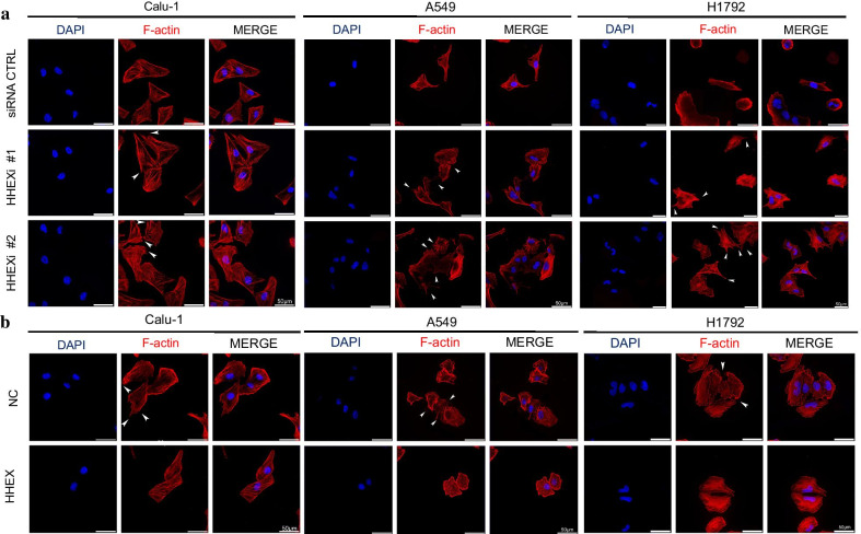
Fig. 2.
Hhex repressed cell protrusion formation. a Calu-1, A549, H1792 cells were transfected with control siRNA or HHEX siRNA. After 24 h, cells were reseeded onto adhesive microscope slides. When cells adhered to slides completely then were fixed with PHEMO buffer. The nuclei were stained with DAPI (blue) and F-actin was stained with TRITC-conjugated phalloidin (red). Scale bars, 50 μm. b Calu-1, A549, H1792 cells were transfected with pcDNA3.1 or pcDNA3.1-HHEX, and the cells were reseeded onto adhesive microscope slides after 24 h of transfection. F-actin was stained with TRITC-conjugated phalloidin (red), and the nuclei were visualized with DAPI (blue). Arrows stand for the typical protrusions, scale bars, 50 μm

