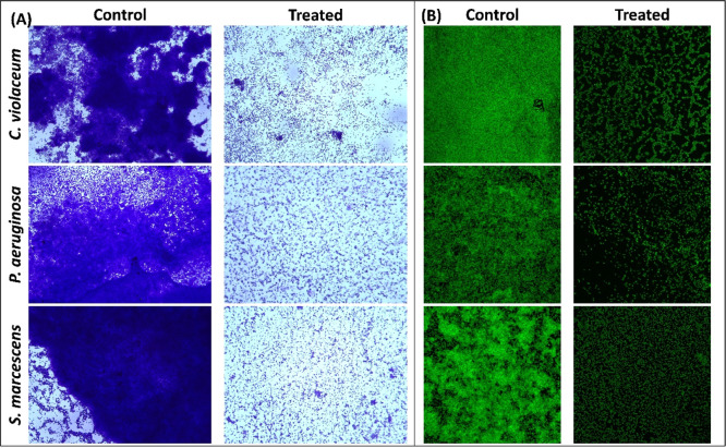
Figure 3.
(A) Light microscopic images of C. violaceum 12472, P. aeruginosa PAO1, and S. marcescens MTCC 97 biofilms in the absence and presence of sub-MIC coumarin. (B) Confocal laser scanning microscopic images of C. violaceum 12472, P. aeruginosa PAO1, and S. marcescens MTCC 97 biofilms in the absence and presence of sub-MIC coumarin. Sub-MIC against C. violaceum 12472 was 100 μg/mL, and sub-MIC against P. aeruginosa PAO1 and S. marcescens MTCC 97 was 250 μg/mL.

