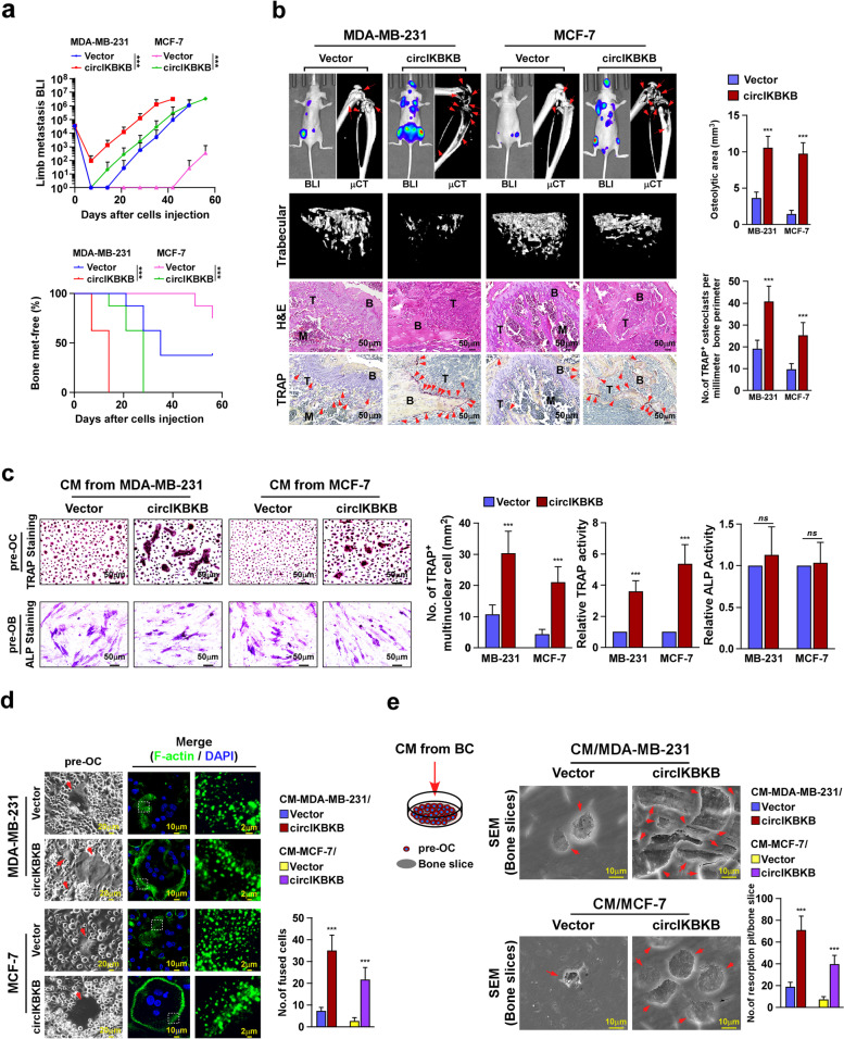
Fig. 2.
Overexpression of circIKBKB promotes osteolytic bone metastasis of breast cancer in vivo. a Normalized BLI signals of bone metastases and Kaplan–Meier bone metastasis-free survival curve of mice from the indicated experimental groups (n = 8/group). b Left: BLI, μCT (longitudinal and trabecular section) and histological (H&E and TRAP staining) images of bone lesions from representative mice. Scale bar, 50 μm. Right: Quantification of μCT osteolytic lesion area and TRAP+ osteoclasts along the bone-tumor interface of metastases from experiment in the left panel. c Osteoclast differentiation assay by TRAP staining (upper) or osteoblast differentiation assay by ALP staining (lower) in the presence of CM from indicated cells. Right: Quantification of number of TRAP+-multinuclear osteoclasts, TRAP activity and ALP activity from experiment in left panel. d Left: Phase contrast micrograph of pre-osteoclasts treated with CM from indicated cells (left) and IF staining images of phalloidin (F-actin) (middle and right). Scale Bar, 20 µm (left), 10 µm (middle) and 2 µm (right). Right: Quantification of the number of fused multinuclear cells from experiment in the left panel. e Bone resorption assay analysis of pre-osteoclasts cultured onto the bone slices treated with CM from indicated cells (left), then bone slice was fixed for scanning electron microscopy (SEM) (middle) and quantification of the number of resorption pits per bone slice (right). Each error bar represents the mean ± SD of three independent experiments. * P < 0.05, ** P < 0.01, *** P < 0.001

