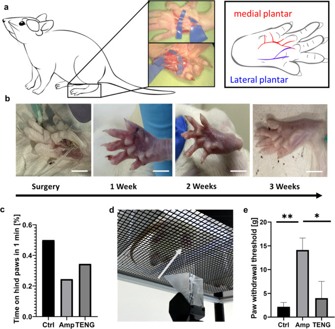
Figure 5.
a. Schematic illustration based on anatomical dissection of the nerves of a Wistar rat’s hindfoot. b. Images of surgical implantation of TENG-IT (8 mm × 3 mm) in a rat’s left hindfoot, and post-operative recovery (scale = 1 cm). c. Percentage of time spent on hind paws for each group, over the course of 1 min. d. von Frey test setup. Arrow points to the tip of the device. e. Difference in average required peak force between the right and left hind paws in the von Frey test, measured for three experimental groups: control, amputee, TENG-IT (P-values: **p = 0.0099, *p = 0.0242).

