Figure 5.
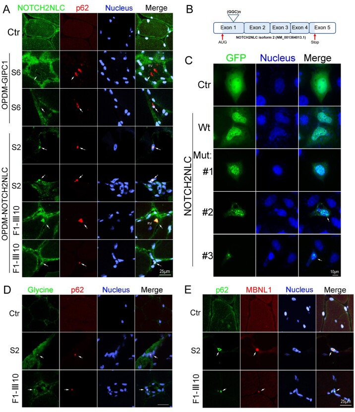
Immunofluorescence on muscle biopsy samples and expression of NOTCH2NLC wild-type or mutant in HEK293 cells. (A) NOTCH2NLC distribution in skeletal muscle of control and OPDM patients. Immunofluorescence showing NOTCH2NLC and p62 co-localized in intranuclear inclusions and rimmed vacuoles of OPDM3 patients (Patients S2 and F1-III10), but not in control or GIPC1-affected OPDM patient (Patient S6), indicated by arrows. (B) Location of GGC repeat expansions in the coding region in the transcript isoform 2 (NM_001364013.1). (C) HEK293 cells were transfected with the control (GFP), NOTCH2NLC wild-type [NOTCH2NLC-(GGC)9] or mutant [NOTCH2NLC-(GGC)69] vectors and subjected to fluorescence observation at 48 h post-transfection. NOTCH2NLC wild-type (Wt) was primarily localized in the nucleus compared to the control, whereas the NOTCH2NLC mutant formed protein aggregates in the nucleus. Different stages of NOTCH2NLC-(GGC)69 aggregation were shown: diffuse small aggregates in Mutant 1, large aggregates in Mutants 2 and 3 (indicated by arrows). The apoptotic cell with condensed nuclei is indicated in Mutant 3. (D) Glycine and p62 co-localized in intranuclear inclusions of OPDM3 patients (Patients S2 and F1-III10), but not in the control (indicated by arrows). (E) RNA binding protein MBNL1 form aggregates in the intranuclear inclusions in OPDM3 patients. Scale bars = 25 μm (A, D and E), and 10 μm (C). Nuclei were counterstained with DAPI.

