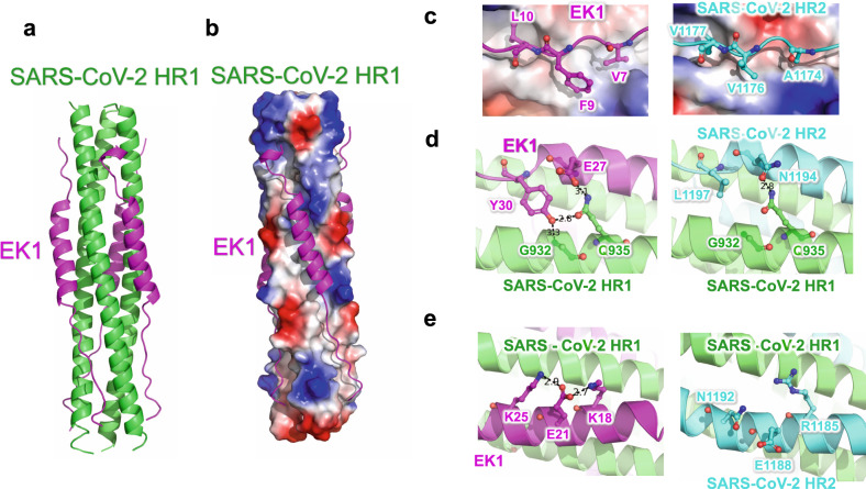
Fig. 4.
Crystal structure of 6-HB formed by SARS-CoV-2-HR1 and EK1. a The structure of SARS-CoV-2-HR1 and EK1 is shown in cartoon representation with HR1 colored in green and EK1 in magenta. b HR1-trimer of SARS-CoV-2 is shown in electrostatic surface, and EK1 is shown in cartoon representation. c–e Interactions of SARS-CoV-2 HR1 motif against EK1 peptide and SARS-CoV-2 HR2 motif. Important binding residues are shown in sticks and labeled

