
Figure 1.
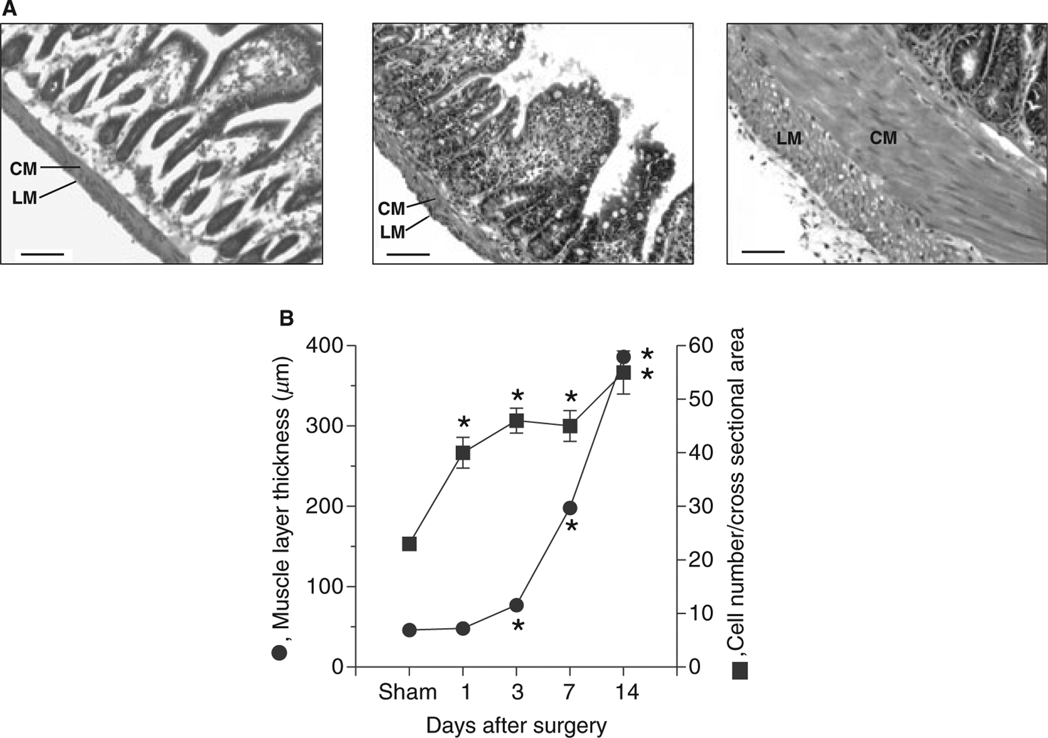
Morphological changes of smooth muscles following partial intestinal obstruction. (A) Representative haematoxylin and eosin-stained sections of sham-operated (left panel), 3 days (middle panel), and 14 days post-obstruction small intestine (right panel) (scale bar: 100 μm) (n = 5). Sections were obtained from sham-operated animals 7 days after sham surgery. No differences were noted in the muscle layers from sham-operated animals 1, 3, 7 and 14 days after sham-surgery (data not shown). CM, circular muscle layer; LM, longitudinal muscle layer; (B) Changes in circular muscle layer thickness and smooth muscle cell number following partial intestinal obstruction (data obtained from four different points from five sham-operated and five small intestine partial-obstruction animals). The number of smooth muscle cells was evaluated by counting the number of nuclei in a 100 μm cross-section of the circular muscle layer (mean ± SE, *P < 0.05 compared with sham).