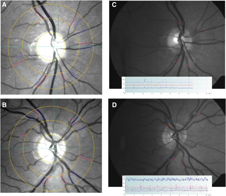
Figure 1. Measurement of static and dynamic retinal vessel diameter.
(A) and (B) Semiautomated measurement of retinal vessel caliber from fundus photos using the program VesselMap2. Measurements are taken from vessels passing through an annular zone 0.5 to 1 disc diameter away from the margin. (C) and (D) Measurement of retinal vessel pulsation amplitudes from video fundoscopy using the DVA.

