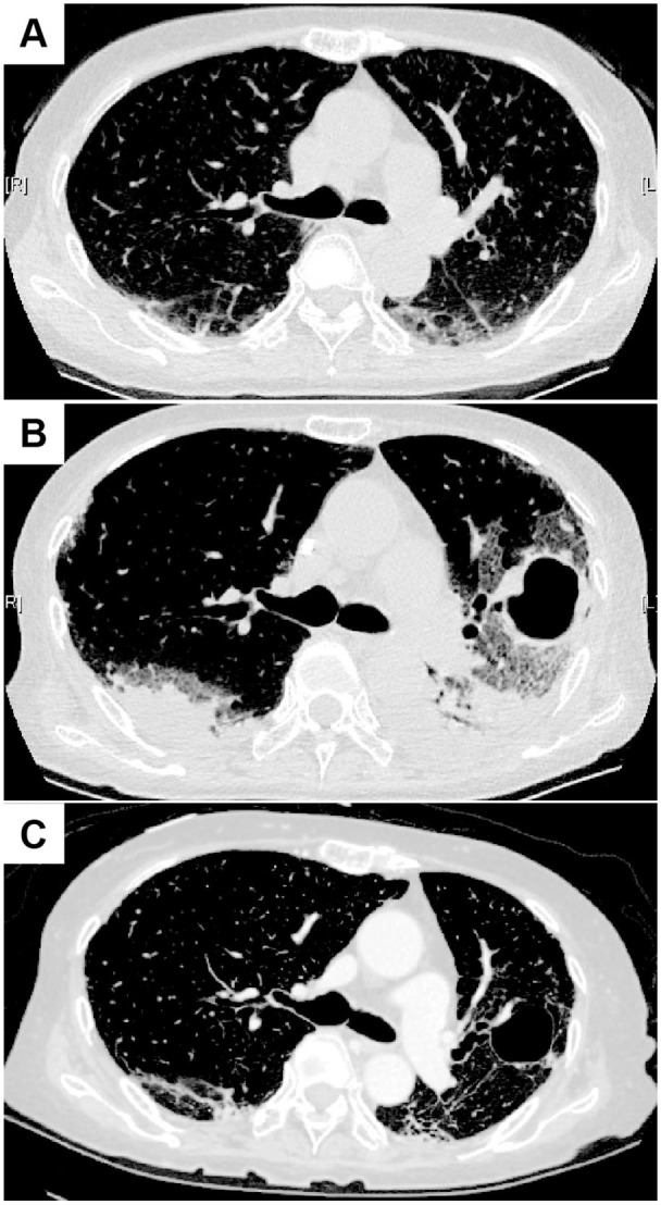
Figure 1.

Chest CT images in Case 1. (A) At the admission. Ground glass opacity and reticular shadows on the peripheral and basilar regions of both lungs were seen. (B) Immediately after the start of the first PE session. Invasive shadows on the dorsal side of the both lungs were seen. In addition, a large cavity with wall thickening was seen in the left lung. (C) After the termination of PE sessions. The ground glass opacity on both lungs still remained, but improved.
