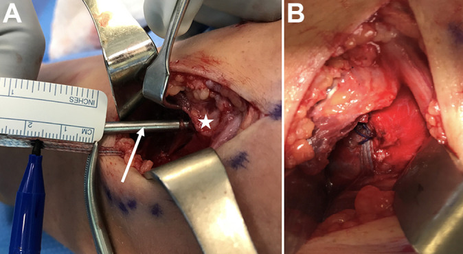
Figure 4.

A right elbow is shown in both images. (A) The first anchor has already been inserted into the sublime tubercle (left side), and the white star marks the medial epicondyle. After drilling and tapping (white arrow) for the second suture anchor, the internal brace is pulled to its desired tension and marked with a hemostat at the level of the bone. The tape is marked 15.5 mm away from the hemostat. The anchor is loaded to this mark and inserted. This compensates for the length of the anchor and avoids overtensioning the internal brace. (B) The final construct after backup suture fixation with a 2-0 braided suture.
