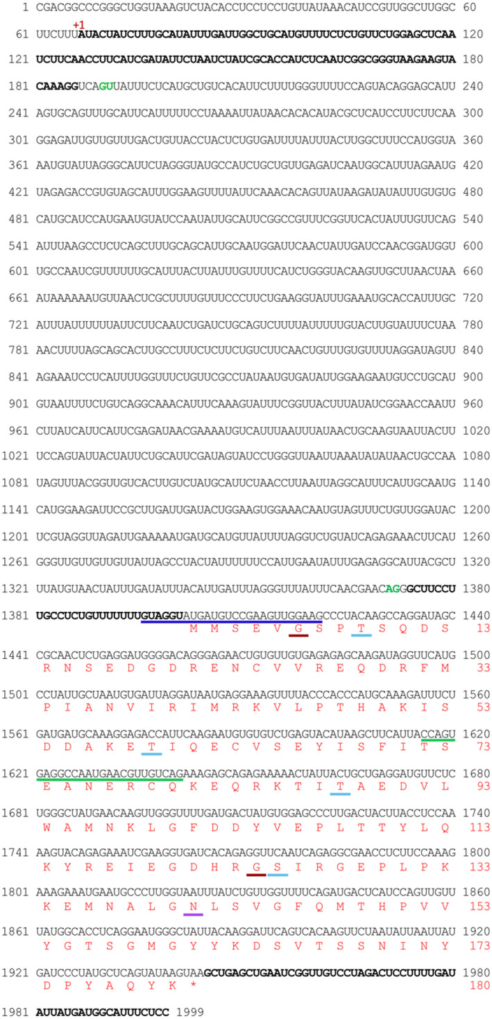
FIGURE 9.

Complete PmLEC1 nucleotide sequence, 5′ UTR intron and deduced protein sequence with putative sites for post‐translational modifications. The nucleotide sequence upstream of the coding region was obtained by genome walking. The green line corresponds to the complementary DNA sequence and represents GSP1, employed in the primary PCR reaction of genome walking, while the blue line corresponds to the complementary DNA sequence and represents GSP2 employed in the secondary PCR reaction. The 5′ UTR and 3′ UTR sequences are displayed in bold. The 5′ UTR is split into two sections by the 5′ UTR intron. The potential intron splice sites are in green. The deduced protein sequence is shown in red and numbered on the right. Putative sites of post‐translational modifications are underlined amino acid residues: dark‐green, N‐myristoylation; light‐blue, phosphorylation; violet, N‐glycosylation
