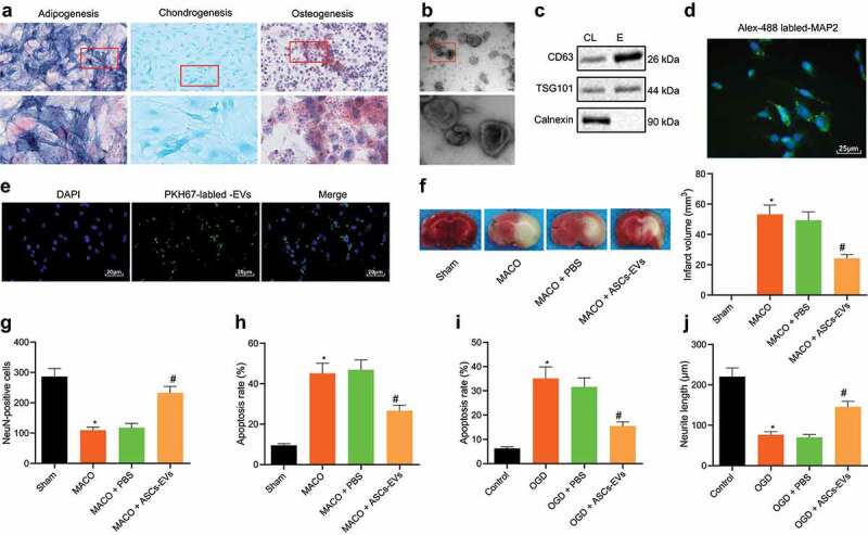
Figure 1.

ASC-EVs protect neuronal cells against cerebral ischaemia/reperfusion
A: Osteogenic, adipogenic and chondrogenic differentiation of ASCs determined by ALP (200 ×), Oil red O staining (400 ×), and Alcain blue staining (400 ×), respectively. B: Observation of EV morphology under a TEM. Scale bar = 100 nm. C: Western blot analysis of EV specific surface marker proteins in EVs. D: Immunofluorescence analysis of MAP2 expression in mouse primary cortical neuronal cells (400 ×). E: Fluorescence microscope analysis of the internalization of EVs by neuronal cells following 24 h of incubation of PKH67-labelled EVs with neuronal cells (400 ×). F: TTC staining of cerebral infarct area in brain tissues of MCAO mice treated with ASC-EVs. G: NeuN immunofluorescence staining of living neuronal cells in brain tissues of MCAO mice treated with ASC-EVs. H: TUNEL staining of cell apoptosis in MCAO mouse brain tissues. I: Flow cytometric analysis of apoptosis of OGD-induced neuronal cells treated with ASC-EVs. J: The neurite outgrowth length of OGD-induced neuronal cells treated with ASC-EVs. *, p < 0.05, vs. the sham-operated mice or control cells; #, p < 0.05, vs. MACO mice treated with PBS or OGD-induced neuronal cells treated with PBS. n = 8 for mice in each group. The measurement data were expressed as mean ± standard deviation of at least three samples. Data comparison among multiple groups was performed using one-way ANOVA and Tukey’s post hoc test. Cell experiments were repeated three times independently.
