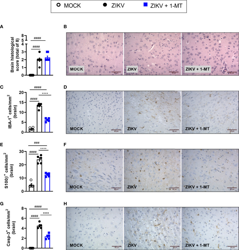
Figure 4.
Morphometric analysis of the brain of A129 mice infected by ZIKV, treated or not with 1-MT inhibitor. A129 were inoculated (i.v) with 4x10³ PFU/200μL of ZIKV and treated daily with 1-MT. (A) Semiquantitative analysis (histopathological score) after H&E staining of brain sections of ZIKV-infected mice 5 days after infection. (C, E, G) Immunostaining of (C) IBA-1+, (E) S100-β+ and (G) Caspase-3 was performed in the brains of mice. (B, D, F, H) Representative images from brain sections. All results are expressed as mean and error bar indicate the standard error (SEM) and are representative of at least two independent experiments. Statistically significant differences were assessed by One-way ANOVA plus Tukey’s comparisons test. (###) for P < 0,001 and (####) for P < 0,0001 when compared to the MOCK group. (****) for P < 0,0001 compared to the ZIKV group.

