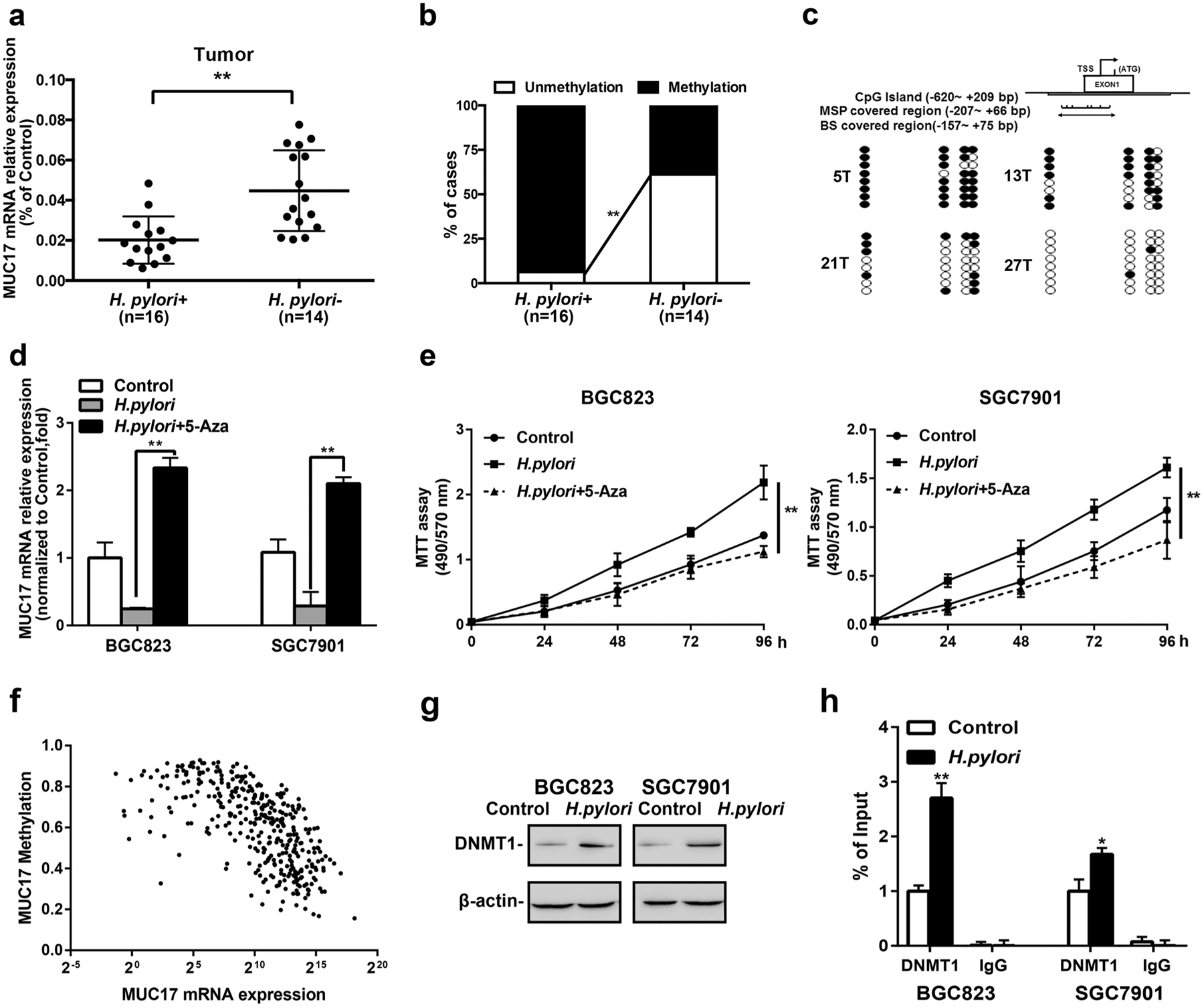
Fig. 2.

Epigenetic silencing of MUC17 by H. pylori in GC. a RT-qRCR of MUC17 expression in human GC tissues with respect to H. pylori infection (n = 30). b Methylated PCR analysis of the promoter methylation of MUC17 in the human GC tissues upon H. pylori infection (n = 30). c BS confirmation of MUC17 gene methylation. Representative GC tissue samples from Hp+ (5T and 13T) or Hp− (21T and 27T). Filled circles represent methylated CpG dinucleotides, and open circles indicate unmethylated sites. d GC cell lines with H. pylori infection were treated with 5-Aza (5 μM, 96 h). MUC17 mRNA expression, relative to β-actin as internal control, was measured by quantitative RT-PCR using the 2−ΔΔCt method. Results represent the mean ± SD (standard deviation). *p < 0.05, **p < 0.01 (by t test) compared with untreated control. e The effects of 5-Aza on GC cell proliferation. MTT assays monitoring the cell variability of H. pylori-infected GC cells treated with 5 μM 5-Aza daily for 3 days. Left panel: BGC823. Right panel: SGC7901. f Association between methylation and expression of MUC17 in GC by analyzing TCGA database. g Western blotting of DNMT1 expression in BGC823 and SGC7901 cells after H. pylori infection (MOI 50, 12 h), β-actin served as protein loading control. (h) ChIP-qPCR to analyze the enrichment of the MUC17 promoter region by DNMT1 in BGC823 and SGC7901 cells after H. pylori infection (MOI 50, 12 h). The specific enrichment was normalized to nonspecific control and was represented as the percentage of input. Data are representative of at least three independent experiments. Error bars indicate standard deviation (SD). *p < 0.05, **p < 0.01, significant differences from the control
