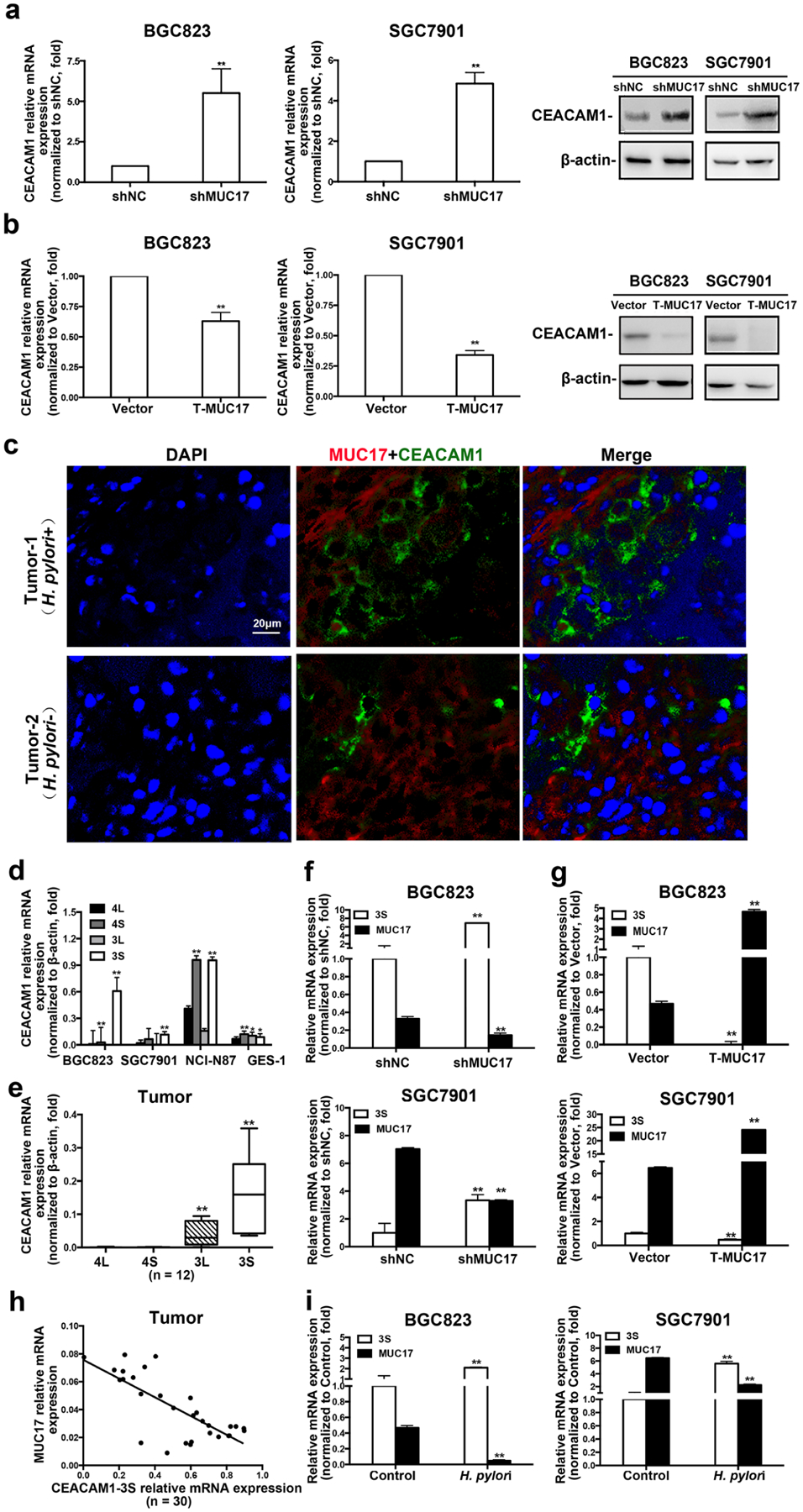
Fig. 4.

MUC17 downregulated the expression of CEACAM1-3S in human GC cells and tissues. a, b RT-qRCR and Western blot analysis of CEACAM1 expression in BGC823 and SGC7901 cells with knockdown of MUC17 (a) or ectopic expression of T-MUC17 (b). c Immunofluorescence staining of MUC17 (red) and CEACAM1 (green) in GC tissues. Nuclei shown by DAPI staining (blue). Shown are results from one of two comparable experiments. Scale bars, 20 μM. d RT-qRCR of the expression levels of CEACAM1 variants (CEACAM1-4L, -4S, -3L, and -3S) in BGC823, SGC7901, NCI-N87 and GES-1 cells. e RT-qRCR of the expression of CEACAM1 variants in GC tissues (n = 12). Analysis of CEACAM1-3S expression in BGC823 and SGC7901 cells by RT-qRCR after knockdown of MUC17 (f) or ectopic expression of T-MUC17 (g). h Correlation between mRNA of MUC17 and CEACAM1-3S in human GC tissues determined by RT-qPCR o (n = 30, p < 0.001, r = − 0.6282). i RT-qPCR of the expression of MUC17 and CEACAM1-3S in GC cells with H. pylori infection (MOI 50,12 h)
