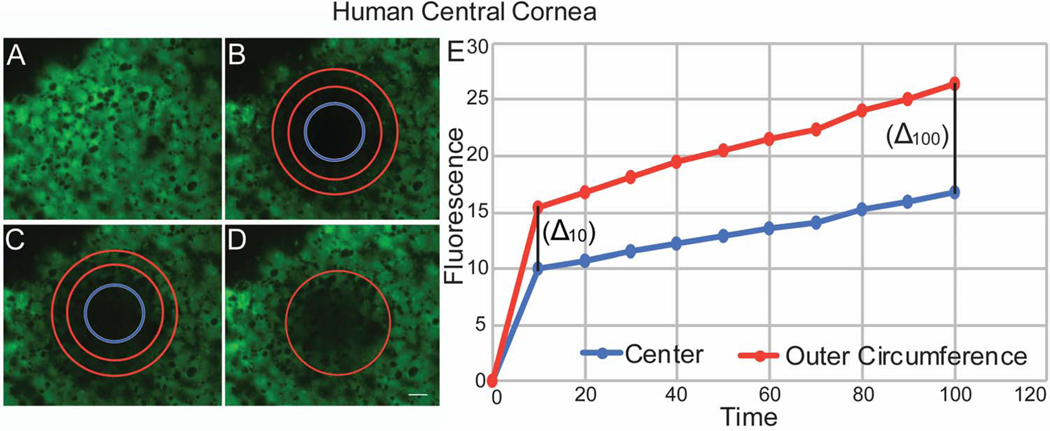
Figure 6. Endothelial – Endothelial Communications Demonstrated by Fluorescence Recovery in the Human Central Cornea after photo-bleaching.
(A) Human endothelial cells were uploaded with calcein AM. (B) A circular area of the endothelial monolayer was photo-bleached. (C) The intensity of fluorescence recovery after photobleaching was evaluated at the 2 different areas of the circle. (D) Endothelial cells recovered fluorescence at 100 seconds. (E) Graphic shows speed and pattern of fluorescence recovery in the central cornea similar to murine. Note that endothelial cells in the outer circumference of the bleached circle recover fluorescence first, followed by the central endothelial cells. This image does not represent table 2.

